
 Login/Register
 Supplier Login


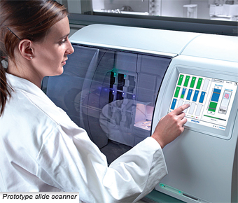 COLLABORATION is in the works to develop quick and reliable digital pathological technology aimed at improving productivity and diagnostic reliability of pathological departments. Royal Philips Electronics and Dako, a Danish company specialising in tissue-based cancer diagnostics, recently agreed to integrate a selection of Dako's image analysis applications into Philips' future digital pathology solutions. A fast pathology slide scanner and an associated image management system form the basis of Philips' proposed integrated solutions for digitising pathology workflows. "Anatomic pathology is an essential element of virtually every cancer diagnosis and the demand for it is ever-increasing. Our goal is to develop integrated digital solutions that enhance the operational efficiency and productivity of pathology departments as well as increasing diagnostic confidence," says Bob van Gemen, general manager, Philips Digital Pathology. The Philips-Dako collaboration will initially focus on leveraging Dako's image analysis software for tissue-based breast cancer diagnosis using its reagents for staining HER2, estrogen receptor, progesterone receptor, p53 and Ki-67 proteins. The detection and quantification of these proteins in biopsy tissue are highly relevant for the classification of breast cancers and the selection of appropriate therapy. "By joining forces with Philips, we will be able to deliver highly competitive diagnostic tools based on Philips' extensive clinical expertise and technology know-how and Dako's expertise in advanced staining and image analysis in order to benefit pathology laboratories, pathologists and ultimately patients," says Lars Holmkvist, CEO of Dako. "I am convinced that our partnership with Dako, with its expert knowledge in detecting and quantifying specific biomarkers in cancer tissue, will significantly accelerate our clinical applications development programme," van Gemen adds. Technology Whose Time Has Come Digitising the images that pathologists normally view through a microscope may enable the introduction of objective and quantitative image analysis tools. Currently, anatomic pathology workflows to examine tissue samples are based on the microscope, through which pathologists examine tissue sections mounted on glass slides and treated with different stains. The staining enhances the contrast between, or reveals the presence of, cellular and molecular components, such as cell nuclei or specific proteins. Accurate interpretation of the results is critical to the diagnosis and staging of each individual patient's disease and requires a great deal of skill and experience. Digitising the images that pathologists normally view through a microscope may enable the introduction of objective and quantitative image analysis tools. Clive Taylor, MD, PhD, professor at University of Southern California and a renowned expert in pathology, expresses about the collaboration: "Digital pathology has been long in gestation, in comparison to radiology, where images also are the currency of practice, and where image acquisition, transfer, interpretation and storage are almost entirely digital. In part, this lag is because acquisition of histopathology images is dependent upon a 100-year-old technique of 'tissue fixation,' sectioning and staining. In part, it is because, somewhat surprisingly, fully digitised histopathology images are much larger than CT files and difficult to manage and analyse. Progress has been slow because there has been no single institution, or company, that embraces both of these areas. It is exciting that collaborations like that between Dako and Philips are now bringing diverse but appropriate expertise to bear on implementing a full digital pathology programme." Philips and Dako will also explore the possibility of extending the collaboration to include image analysis software for immunohistology-basNike Zoom Live 2017
Joining Forces in Digital Pathology
Source: Release Date:2010-10-09 450
Share to: 


COLLABORATION is in the works to develop quick and reliable digital pathological technology aimed at improving productivity and diagnostic reliability of pathological departments. Royal Philips Electronics and Dako, a Danish company specialising in tissue-based cancer diagnostics, recently agreed to integrate a selection of Dako's image analysis applications into Philips' future digital pathology solutions. A fast pathology slide scanner and an associated image management system form the basis of Philips' proposed integrated solutions for digitising pathology workflows. "Anatomic pathology is an essential element of virtually every cancer diagnosis and the demand for it is ever-increasing. Our goal is to develop integrated digital solutions that enhance the operational efficiency and productivity of pathology departments as well as increasing diagnostic confidence," says Bob van Gemen, general manager, Philips Digital Pathology. The Philips-Dako collaboration will initially focus on leveraging Dako's image analysis software for tissue-based breast cancer diagnosis using its reagents for staining HER2, estrogen receptor, progesterone receptor, p53 and Ki-67 proteins. The detection and quantification of these proteins in biopsy tissue are highly relevant for the classification of breast cancers and the selection of appropriate therapy. "By joining forces with Philips, we will be able to deliver highly competitive diagnostic tools based on Philips' extensive clinical expertise and technology know-how and Dako's expertise in advanced staining and image analysis in order to benefit pathology laboratories, pathologists and ultimately patients," says Lars Holmkvist, CEO of Dako. "I am convinced that our partnership with Dako, with its expert knowledge in detecting and quantifying specific biomarkers in cancer tissue, will significantly accelerate our clinical applications development programme," van Gemen adds. Technology Whose Time Has Come Digitising the images that pathologists normally view through a microscope may enable the introduction of objective and quantitative image analysis tools. Currently, anatomic pathology workflows to examine tissue samples are based on the microscope, through which pathologists examine tissue sections mounted on glass slides and treated with different stains. The staining enhances the contrast between, or reveals the presence of, cellular and molecular components, such as cell nuclei or specific proteins. Accurate interpretation of the results is critical to the diagnosis and staging of each individual patient's disease and requires a great deal of skill and experience. Digitising the images that pathologists normally view through a microscope may enable the introduction of objective and quantitative image analysis tools. Clive Taylor, MD, PhD, professor at University of Southern California and a renowned expert in pathology, expresses about the collaboration: "Digital pathology has been long in gestation, in comparison to radiology, where images also are the currency of practice, and where image acquisition, transfer, interpretation and storage are almost entirely digital. In part, this lag is because acquisition of histopathology images is dependent upon a 100-year-old technique of 'tissue fixation,' sectioning and staining. In part, it is because, somewhat surprisingly, fully digitised histopathology images are much larger than CT files and difficult to manage and analyse. Progress has been slow because there has been no single institution, or company, that embraces both of these areas. It is exciting that collaborations like that between Dako and Philips are now bringing diverse but appropriate expertise to bear on implementing a full digital pathology programme." Philips and Dako will also explore the possibility of extending the collaboration to include image analysis software for immunohistology-basNike Zoom Live 2017Share to:
You May Like