
 Login/Register
 Supplier Login


 At the press conference, MIH was appointed by Synthes, a leading global medical device company, to be the first training centre in Asia Pacific for the latest technique. Dr. Hans Schrader, Executive Medical Director of MIH, talked about MIH's move to become the Centre of Excellence in orthopaedics and spinal surgery. Michael Mayer, Professor of Neurosurgery at Salzburg Medical School and the Medical Director & Head of Spine and Orthopaedic Centre Munich, together with Dr. Clarence Leung, Specialist in Neurosurgery, shared more details about this advanced technique and its application in Hong Kong. What is cervical artificial disc replacement surgery? Cervical artificial disc replacement surgery is a procedure to replace the prolapsed or herniated intervertebral disc between each pair of vertebrae in the neck. The key feature is the insertion of an artificial disc between the vertebrae to replace the natural spinal disc after it has been removed for reasons such as degeneration of the discs caused by age, genetics or daily wear and tear. Use of sophisticated materials for improved MRI compatibility The new artificial disc is composed of top and bottom endplates and a plastic inlay that forms a ball-and-socket joint. This design enables motion by allowing the top endplate to move over the plastic ball attached to the bottom endplate. Instead of metal components, the top and bottom plates are made of titanium, so the new implant is MRI compatible, providing the surgeon with high-quality diagnostic information. The ball-and-socket is made of cobalt chrome and polyethylene for durability to bear the wear-and-tear motion in the joint. Special keel design for enhancing multilevel replacement The anchoring keels on the endplates come with a new tripod design and smaller dimensions. There is a central keel on the top endplate and two smaller keels on the lower plate. The new configuration enhances the stability in multilevel replacement and minimises the "stress" of adjacent keels in multilevel procedures. Several new instruments have also been introduced to improve the precision of the procedure, including positioning, removal of the herniated disc, defining the size of the implant and insertion of the artificial disc. The precision-cutting system makes use of an electric cutter, which makes keel cuts smoothly and reduces the impact on bone and spinal cord. Generally, the operation takes about 1 to 2 hours. In normal circumstances, the patient can be mobilised and discharged the day after the surgery compared to 1 to 2 weeks for patients having a fusion procedure. Who are the candidates for this procedure? The new technique offers an alternative option to young patients who did not respond to nonoperative care for cervical degenerative diseases, trapped nerves, spinal cord compression and severe neck pain. It is estimated that 30% to 40% of the Hong Kong population above the age of 40 have cervicaPW Human Race NMD
The Latest Cervical Artificial Disc Replacement Technique
Source: Release Date:2010-07-27 642
Share to: 


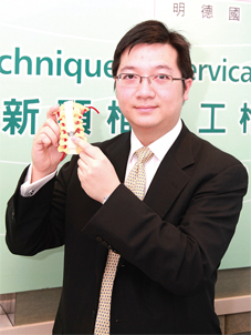
HONG KONG-Having introduced several leading spinal and orthopaedic procedures in Hong Kong, Matilda International Hospital (MIH) again introduced the latest technique in cervical artificial disc replacement surgery in a recent press conference held last June 19, 2010 at 4/F, Lecture Hall, 41 Mount Kellett Road, The Peak. With the help of special precision instruments, surgeons replace patients' prolapsed cervical disc with a cutting-edge artificial disc in an anterior approach (from the front of the neck). The artificial disc is made of sophisticated materials and comes with a special implant design that improves the magnetic resonance imaging (MRI) compatibility and enhances the stability in multilevel disc replacement. This innovative technique offers an alternative option to patients who require a surgery for cervical disc degenerative diseases. "Alternative option to patients who require a surgery for cervical disc degenerative diseases" At the press conference, MIH was appointed by Synthes, a leading global medical device company, to be the first training centre in Asia Pacific for the latest technique. Dr. Hans Schrader, Executive Medical Director of MIH, talked about MIH's move to become the Centre of Excellence in orthopaedics and spinal surgery. Michael Mayer, Professor of Neurosurgery at Salzburg Medical School and the Medical Director & Head of Spine and Orthopaedic Centre Munich, together with Dr. Clarence Leung, Specialist in Neurosurgery, shared more details about this advanced technique and its application in Hong Kong. What is cervical artificial disc replacement surgery? Cervical artificial disc replacement surgery is a procedure to replace the prolapsed or herniated intervertebral disc between each pair of vertebrae in the neck. The key feature is the insertion of an artificial disc between the vertebrae to replace the natural spinal disc after it has been removed for reasons such as degeneration of the discs caused by age, genetics or daily wear and tear. Use of sophisticated materials for improved MRI compatibility The new artificial disc is composed of top and bottom endplates and a plastic inlay that forms a ball-and-socket joint. This design enables motion by allowing the top endplate to move over the plastic ball attached to the bottom endplate. Instead of metal components, the top and bottom plates are made of titanium, so the new implant is MRI compatible, providing the surgeon with high-quality diagnostic information. The ball-and-socket is made of cobalt chrome and polyethylene for durability to bear the wear-and-tear motion in the joint. Special keel design for enhancing multilevel replacement The anchoring keels on the endplates come with a new tripod design and smaller dimensions. There is a central keel on the top endplate and two smaller keels on the lower plate. The new configuration enhances the stability in multilevel replacement and minimises the "stress" of adjacent keels in multilevel procedures. Several new instruments have also been introduced to improve the precision of the procedure, including positioning, removal of the herniated disc, defining the size of the implant and insertion of the artificial disc. The precision-cutting system makes use of an electric cutter, which makes keel cuts smoothly and reduces the impact on bone and spinal cord. Generally, the operation takes about 1 to 2 hours. In normal circumstances, the patient can be mobilised and discharged the day after the surgery compared to 1 to 2 weeks for patients having a fusion procedure. Who are the candidates for this procedure? The new technique offers an alternative option to young patients who did not respond to nonoperative care for cervical degenerative diseases, trapped nerves, spinal cord compression and severe neck pain. It is estimated that 30% to 40% of the Hong Kong population above the age of 40 have cervicaPW Human Race NMD
At the press conference, MIH was appointed by Synthes, a leading global medical device company, to be the first training centre in Asia Pacific for the latest technique. Dr. Hans Schrader, Executive Medical Director of MIH, talked about MIH's move to become the Centre of Excellence in orthopaedics and spinal surgery. Michael Mayer, Professor of Neurosurgery at Salzburg Medical School and the Medical Director & Head of Spine and Orthopaedic Centre Munich, together with Dr. Clarence Leung, Specialist in Neurosurgery, shared more details about this advanced technique and its application in Hong Kong. What is cervical artificial disc replacement surgery? Cervical artificial disc replacement surgery is a procedure to replace the prolapsed or herniated intervertebral disc between each pair of vertebrae in the neck. The key feature is the insertion of an artificial disc between the vertebrae to replace the natural spinal disc after it has been removed for reasons such as degeneration of the discs caused by age, genetics or daily wear and tear. Use of sophisticated materials for improved MRI compatibility The new artificial disc is composed of top and bottom endplates and a plastic inlay that forms a ball-and-socket joint. This design enables motion by allowing the top endplate to move over the plastic ball attached to the bottom endplate. Instead of metal components, the top and bottom plates are made of titanium, so the new implant is MRI compatible, providing the surgeon with high-quality diagnostic information. The ball-and-socket is made of cobalt chrome and polyethylene for durability to bear the wear-and-tear motion in the joint. Special keel design for enhancing multilevel replacement The anchoring keels on the endplates come with a new tripod design and smaller dimensions. There is a central keel on the top endplate and two smaller keels on the lower plate. The new configuration enhances the stability in multilevel replacement and minimises the "stress" of adjacent keels in multilevel procedures. Several new instruments have also been introduced to improve the precision of the procedure, including positioning, removal of the herniated disc, defining the size of the implant and insertion of the artificial disc. The precision-cutting system makes use of an electric cutter, which makes keel cuts smoothly and reduces the impact on bone and spinal cord. Generally, the operation takes about 1 to 2 hours. In normal circumstances, the patient can be mobilised and discharged the day after the surgery compared to 1 to 2 weeks for patients having a fusion procedure. Who are the candidates for this procedure? The new technique offers an alternative option to young patients who did not respond to nonoperative care for cervical degenerative diseases, trapped nerves, spinal cord compression and severe neck pain. It is estimated that 30% to 40% of the Hong Kong population above the age of 40 have cervicaPW Human Race NMDShare to:
You May Like