
 Login/Register
 Supplier Login


A breakthrough 3D printed airway splint technology saved the lives of three babies with a life-threatening condition, suggesting potential future use for 3D printing even for medical applications.
The results of the groundbreaking procedure, recently published on Science Translational Medicine(*), also indicate early treatment of tracheobronchomalacia may prevent complications of conventional treatment. Kaiba Giofriddo, Garett Peterson and Ian Orbich suffered from a terminal form of tracheobronchomalacia, which causes the windpipe to periodically collapse and prevents normal breathing. There was no cure and life expectancies were grim. Kaiba turned blue when he was just a newborn because his lungs were not getting any oxygen. Garrett spent the first year of his life in hospital beds tethered to a ventilator and fed through his veins because his body was too sick to absorb food. Baby Ian’s heart stopped before he was even six months old.
Kaiba, Garrett and Ian became the first in the world to benefit from specially designed splints aimed at keeping their airways open and restore their breathing.
The bioresorable splints were created by Glenn Green, M.D., associate professional of pediatric otolaryngology at University Michigan’s C.S. Mott Children’s Hospital and his colleague Scott Hollister, Ph.D., professor of biomedical engineering and mechanical engineering and associate professor of surgery at University of Michigan (U-M), using 3D printing.
Doctors Green and Hollister integrated an image-based computer model with laser-based 3D printing to produce the splint based on CT scans of the boy’s tracheas.
Kaiba, the first of the boys to undergo the procedure in 2012 when he was only 3 months old, made national headlines after the treatment. The procedure, which was done at the C.S. Mott Children’s Hospital, was then repeated with Garrett, then 16-months old, Ian, 5-months old.
Doctors received emergency clearance from the FDA to implant the customized tracheal splint for each patient. It involved sewing the splint around each of the boy’s airways to expand the trachea and bronchus and give it a skeleton to aid proper growth.
The splint is designed to be reabsorbed by the body over time.
Bioresorable splints
Doctors Green and Hollister followed the cases closely to see how well the bioresorable splints implanted in all three patients have worked. None of the devices were shown to cause any complications. Further, CT and MRI scans used to monitor the growth of the airways show the splints were opening up to allow airway growth for all three patients.
Kaiba, now an active 3-year-old, was back at Mott recently for a follow-up appointment. Checks reveal that splint is dissolving as expected. The doctors expect that his trachea will eventually reflect that of his peers with no signs of the tracheobronchomalacia that nearly killed him as a newborn.
The findings reveal improvements in multiple organ systems among all three patients. They were able to come off of ventilators and no longer needed paralytics, narcotics and sedation. They were also relieved of immunodeficiency-causing proteins that prevented them from absorbing food and therefore no longer needed intravenous therapy.
Dr. Green said their team is pleased to find all cases have so far proven to improve the lives of the patients. “These cases broke new ground for us because we were able to use 3D printing to design a device that successfully restored patients’ breathing through a procedure that had never been done before,” he said.
In addition, the researchers’ findings indicate early treatment of tracheobronchomalacia may prevent tracheostomy, prolonged hospitalization, mechanical ventilation, cardiac and respiratory arrest, food malabsorption and discomfort resulting from traditional treatment procedures.
“Before this procedure, babies with severe tracheobronchomalacia had little chance of surviving. Today, our first patient Kaiba is an active, healthy 3-year-old in preschool with a bright future. The device worked better that we could have ever imagined. We have been able to successfully replicate this procedure and have been watching patients closely to see whether the device is doing what it was intended to do. We found that this treatment continues to prove to be a promising option for children facing this life-threatening condition that has no cure,” Dr. Green.
Kaiba, who loves playing with his siblings, recently saw his favorite character Mickey Mouse at Disney World, thanks to the Make-a-Wish Foundation.
Despite noting that the device was not designed for safety and that rare potential complications of the procedure may not yet be evident, Dr. Green stated the potential of 3D-printed medical devices to improve outcomes for patients is clear. “But we need more data to implement this procedure in medical practice,” he added.
Richard G. Ohye, M.D., head of pediatric cardiovascular surgery at C.S. Mott who performed the surgeries, said the cases provide the groundwork for exploring a clinical trial that could help other children with less-severe forms of tracheobronchomalacia in the future.
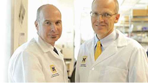
Dr. Scott Hollister and Dr. Glenn Green
* “Mitigation of tracheobronchomalacia with 3D-Printed Personalized Medical Devices in Pediatric Patients, Science Translational Medicine, April 29, 2015